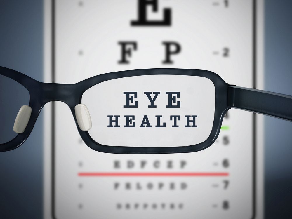
What is an eye-health examination

What is an eye-health examination

What is an eye-health examination

Your experience at our clinic may be different than any other eye exam you have had. Our doctors and staff will go through numerous steps, in painstaking detail, to ensure that we meet the specific needs of each patient.
In order to give you a better understanding of the eye-health examination that you will receive, we would like to “walk you through” an eye examination and explain each step of the process. We want you to be informed about the amount of time required, clinical skills needed, and advanced technology necessary to provide the level of care you will experience at Family Eye Clinic.
Let us begin with the fact that we utilize electronic medical records (EMR). We are proud to say that our clinics are “paperless.” Every aspect of your medical record will be housed electronically, in a secure, encrypted data server. We follow the strict HIPAA privacy guidelines set forth by the department of health and human services, and our software system has met the “meaningful use” criteria implemented by the centers for Medicare services.
A key factor in the quality of an examination is the amount of time the doctors and staff spend with each patient. Our doctors and staff generally spend between one and two hours with you on a comprehensive exam. During your eye-health examination, each of the following procedures provides the doctor with the information necessary to ensure your vision is as clear as possible and your eyes remain healthy.
Exam Process
Patient Information
The first step in an eye health examination is collecting information. Demographic information such as age, race, and occupation, can be very helpful. Certain diseases and eye conditions affect people of different ages and various ethnic backgrounds in different ways. For example, people of African descent are two times as likely to go blind from glaucoma, Hispanics are at a much greater risk for diabetic eye disease, and macular degeneration is the leading cause of blindness in people over 65.
A patient’s medical history is one of the most important elements of an exam. Although it may seem irrelevant, knowing every aspect of your health, as well as any medications you may take, is vital. The eye is not an isolated organ and can be affected by a vast array of systemic conditions and medications. Did you know that diabetes is the leading cause of blindness in the U.S.? Or that there are certain eye drops used to treat glaucoma that cannot be used if you have asthma? Or that inflammatory processes such as arthritis and irritable bowel may cause pain in the eye? This is why having a good medical history is so important. All the systems of the body work together, so your doctor needs to know about any medical condition you may have.
TAKING VITALS
We will take information on your vital signs. These include measuring your height, weight, and blood pressure. These statistics can aide the doctor in assessing your risk for numerous diseases and eye problems. It can also help us ensure that any medications you take are working properly.
LENSOMERTY
We will use an instrument called a digital lensometer to measure your current pair of glasses. This device precisely measures each detail of your glasses prescription, including the amount of correction for nearsightedness, farsightedness, astigmatism, prism, and bifocal power. Having this information will let the doctor know how much change there has been from your current prescription. Knowing this can help the doctor decide if a difference in the glasses prescription is responsible for a change in vision, or if the vision loss may be related to something more serious.
VISUAL ACUITY
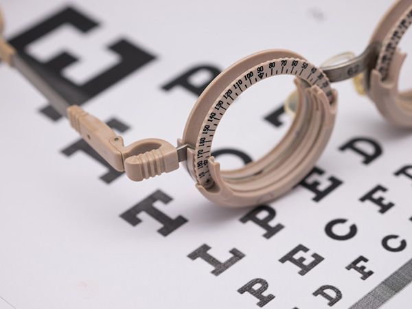
We will measure how well you see both with your current spectacle or contact lenses on and also without correction. Checking the visual acuity will be done at every visit. This can tell the doctor if your current prescription is accurate. It is also one of the most important ways to track the progression or healing of eye diseases, injuries, and infections.
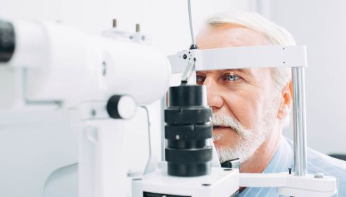
AUTOREFRACTION
An instrument called an OPD Scan will be used to measure the focal length of each eye. Using a system of both infrared and visible light waves this machine measures your optical prescription at over 2,500 points on the surface of the eye. This gives the doctor a very accurate estimation of your prescription needs. It also has the ability to perform wavefront aberrometry, allowing the doctors to detect distortions and aberrations in the optical pathway that have previously been unmeasurable.
KERATOMETRY
This provides detailed information about the curvature of the front surface of the eye, the cornea. Keratometry readings are essential for determining the amount and axis of astigmatism, and the fitting of both rigid (hard) and soft contact lenses. These readings can also help detect and monitor corneal diseases and dystrophies such as keratoconus.
INTRAOCULAR PRESSURE (IOP)
The eye is filled with a fluid called aqueous humor. This fluid exerts a force just as air in a tire or water in a balloon. The pressure of this fluid is critical to the health of the eye. Having IOP’s outside of the normal range may be associated with several abnormal eye conditions. At each eye exam, your IOP will be measured and evaluated by the doctor.
EXTRAOCULAR MOTILITY (EOM)
The movement of each eye is controlled by six muscles. Each muscle has a specific action and is controlled by a specific nerve. These nerves follow intricate routes to control centers in the brain. During your exam, the doctor will test the function of each of the muscles. This allows an assessment of the muscle, the nerve, and also the brain. There are many conditions, including space-occupying lesions such as tumors and aneurysms, which can be detected by testing extraocular motility.
VISUAL FIELDS
The term visual field describes defines the space to the left, right, up, and down from the point of fixation; in other words, your peripheral vision. In order for us to perceive the world, light enters the eye, is absorbed by the retina, converted into an electrical signal, and then sent through the optic nerve and visual pathways of the brain into the occipital cortex. Here it is processed into what we see. This pathway from the eye to the occipital cortex is very structured. By analyzing patterns in visual field loss (missing areas in the peripheral or central vision) it is often possible to identify exactly where along the visual pathway there is a problem. Problems such as strokes, tumors, aneurysms, MS, and increased intracranial pressure, are often discovered and monitored with the help of visual field testing. Your doctor will check the extent of your visual field and if warranted may use very sophisticated equipment to get a complex analysis of it.
PUPILLARY ASSESSMENT
The pupil is the black circle in the center of the colored part (iris) of the eye. It is actually an opening which allows light to enter the eye. This opening gets larger (dilates) in dim light to allow more light into the eye and gets smaller (contracts) in bright light to keep too much light from entering. The neurological pathway for this process is very complex. By testing how your pupils react to light, and how they work with each other, your doctor is able to detect and isolate numerous ocular and neurological problems.
COVER TESTING
While holding your fixation very steady, your doctor will analyze the movements of each eye when covered and then uncovered. Doing this lets the doctor determine many things about the alignment of eyes, the resting positions of each eye, and how well your eyes work together. This procedure can help with detecting and monitoring the progression of several binocular vision abnormalities such as muscle imbalances, eye turns or misalignments, and neurological problems.
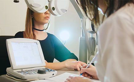
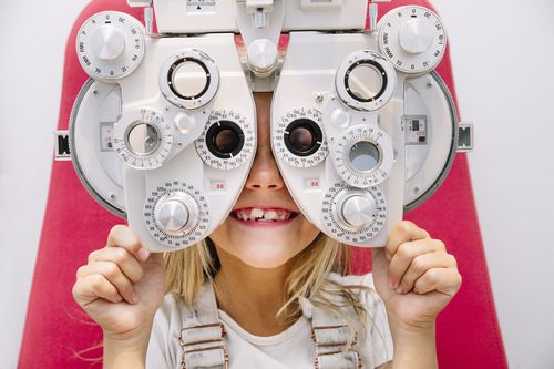
REFRACTION
The refraction is the part of the exam where the doctor determines your optical needs. This is what most people think of when they come to see the eye doctor. Everyone knows the infamous question, “which is better, number 1 or number 2?” As simple as it sounds, by answering this question you give the doctor very critical information. Using your answers the doctor will be able to pinpoint the optical correction that will give you the sharpest vision possible at all distances. During the process, your doctor will also measure details about the alignment of your eyes, how well your eyes work together, and the ability of the eye to change its focus. By knowing these details the doctor will be able to adjust your prescription so that not only is the visual acuity very sharp, but the vision will be comfortable, significantly reducing eye strain and fatigue.
Many times refraction is confused with an eye exam. However, the refraction is only one step in the process of examining the eyes. Although good refraction is a key element, there is much more to a comprehensive eye examination. In fact, most insurances require the refraction to be coded and billed separately from the eye exam. So if you notice this on your statement from our office or your insurance, do not worry.
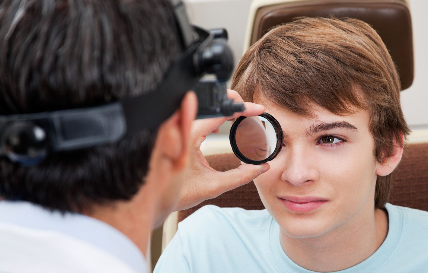
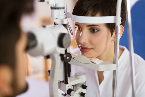
SLIT LAMP EXAMINATION
The eye is a relatively small structure. In order to discern the smallest detail about the anatomy of your eye, the doctor will use an instrument called a slit lamp or biomicroscope. This allows the doctor a highly magnified view to evaluate the health of the ocular tissues. With this high level of magnification, your doctor will examine both the surface of the eye including the cornea, tear film, lids, and lashes, as well as internal ocular structures such as the iris, lens, retina, and optic nerve. Finding and treating many eye diseases would be virtually impossible without this tool. Many of our slit lamps are equipped with cameras capable of capturing both still images and video so that the doctors can show you exactly what they see.
RETINAL EXAMINATION
The inner wall of the eye is lined by a tissue called the retina. It is the job of the retina to absorb light and convert this light into an electrical signal that can be sent along the optic nerve to the brain. There are dozens of diseases and abnormalities that may affect the retina and other internal structures of the eye. In order to find these problems, your doctor will do a thorough examination of your retina and the inside of your eye.
Looking into the eye can be a difficult task. The only way to see into the eye is the same way you see out, through your pupil. However, when a light is shined into the eye for the doctor to see in, the pupil constricts very tightly to a size between two and three millimeters. Imagine trying to look into a room and see every detail of the room’s interior while only looking through the keyhole. But luckily there is a better way. By using drops to dilate the pupil, we dramatically increase the doctor's ability to see into the eye. It would be similar to opening the door to the room and looking in versus merely looking through the keyhole.
Dilating the pupils does have some minor side effects, but using the newest type of drops keeps these to a minimum. After having your eyes dilated you may notice a slight blurring of vision (primarily at near) and a sensitivity to light. These symptoms may last up to four hours but usually, subside within two. For some patients, there may now be an alternative to dilation. Advancements in retinal imaging technology have to lead to the development of the OPTOMAP. This imaging system captures a 200-degree field inside the eye, allowing the doctor to see up to ten times more than a standard retinal photograph.
CONTACT LENS FITTING /EVALUATION
If you are a long time wearer of contact lenses or want to try them for the very first time, the doctor will perform a contact lens evaluation, in addition to your eye exam. During this evaluation the doctor will take several steps to make sure the contact lens is fitting well, the vision is clear, and the surface of the eye is healthy. Contact lens evaluations are broken into five different levels based on the amount of care required.
EYEWEAR SELECTION
After meticulously determining your specific visual needs, your doctor will have one of our certified opticians assist you with your eyewear selection. The primary job of the optician is education. They will educate you on the products available that will best fit your lifestyle. The opticians are trained to help you with the fine details of choosing a frame. The perfect frame must be stylish and functional.
Our opticians are experts in the latest spectacle lens technology. They have had extensive training on the most advanced lenses on the market today. We have partnered with Hoya Labs to bring you the Mystyle Lens. Measurements for this lens are totally custom. The opticians will take detailed measurements of the frame on your face. They will also ask a series of questions about how you use your vision each day (computer, piano, golf, etc.) A computer algorithm then custom designs a lens for your individual situation. The lens is created by a digital “free form” computer controlled lathing process, which provides distortion-free clarity that has never before been possible.
ADDITIONAL DIAGNOSTIC TESTING
During your eye examination, the doctor may detect findings related to the health of your eyes that require further evaluation. It is the doctor’s role to leave no stone unturned when it comes to the health of your eyes. Fortunately, our clinics are equipped with cutting edge diagnostic instruments capable of the early detection of a vast array of eye diseases and conditions. Here we will list some of the equipment we use to ensure you receive the most thorough diagnostic and treatment procedures available.
Contact Form
Contact Us
clinic Hours
- Monday8:00 AM - 6:00 PM
- Tuesday8:00 AM - 6:00 PM
- Wednesday8:00 AM - 6:00 PM
- Thursday8:00 AM - 6:00 PM
- Friday8:00 AM - 6:00 PM
- SaturdayClosed
- SundayClosed
Powered by: